News

A closer look into the molecule
The Werner Siemens Imaging Center partners with the University of Tübingen and the University Hospital Tübingen. Together, they are driving forward research in medical imaging technologies. In 2019, the WSIC’s research focus on tumour therapies became part of Germany’s national Excellence Strategy. The next goal? Developing sustainable cancer therapies.
Since its foundation in 2014, the Werner Siemens Imaging Center (WSIC) in Tübingen has grown into one of the world’s leading research facilities for preclinical imaging and medical imaging techniques; basic research on cell and animal models is the focus at WSIC. Professor Bernd Pichler—holder of the Werner Siemens Foundation Endowed Chair—leads the WSIC team of some 60 researchers who contribute their specialist knowledge from the fields of biology, physics, medicine, chemistry and engineering science. The interdisciplinary approach at WSIC is leading to a better understanding of how illnesses arise, progress and spread through the body. “Our overarching goal is to obtain research findings that will lead to more effective treatments of diseases like cancer, Alzheimer’s or Parkinson’s,” says Bernd Pichler.
Combined PET/MRI systems
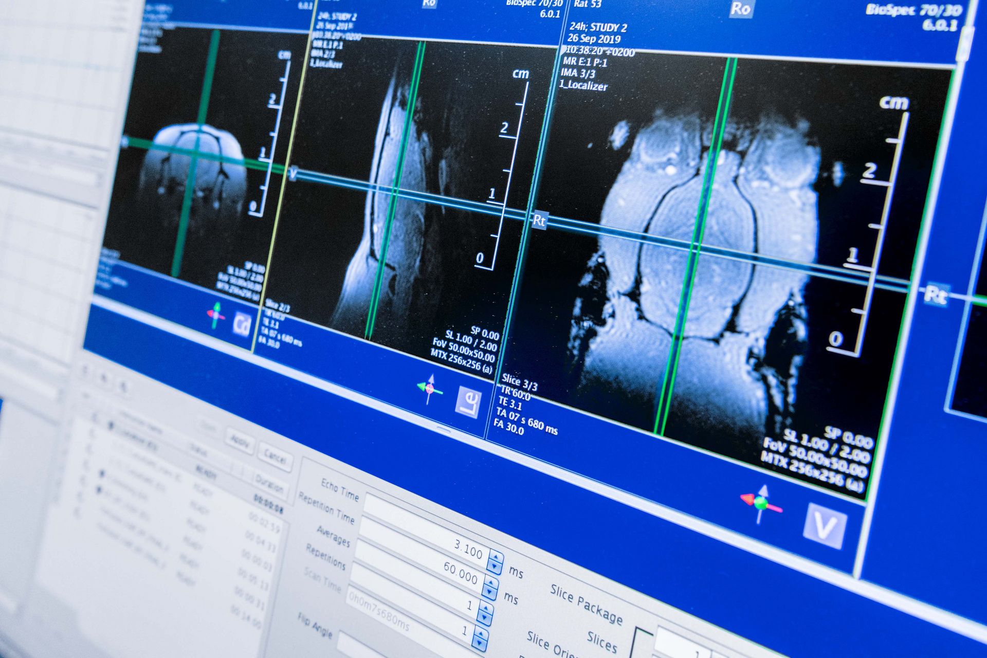
Taking one of the WSIC projects as an example, Bernd Pichler describes the complex research activities he and his team conduct: “At WSIC, we developed a medical imaging system to examine mice. The system combines positron emission tomography with magnetic resonance imaging, and the combined PET/MRI system makes it possible to gain insight into how a disease arises and progresses in the mice’s bodies.” These combined medical imaging systems are of major importance both in research and in cancer diagnostics.
Rapid translation into clinical practice
The team at WSIC is also developing biomarkers: radioactive or fluorescent chemical compounds that medical imaging techniques make visible in the body. For instance, the researchers succeeded in using radioactively labelled antibodies to detect the fungus Aspergillus fumigatus, which is extremely dangerous to humans. To ensure that patients can benefit from the stream of new findings as quickly as possible, the WSIC team works closely with doctors and researchers at the University Hospital Tübingen—and in 2019, the procedure for fungus labelling and diagnostics was successfully used on the first patients at the hospital. A new biomarker is also being developed for early recognition of Parkinson’s symptoms, and Bernd Pichler reports that excellent progress has been made in medical imaging of tumours.
National success
Germany’s national Excellence Strategy promotes state-of-the-art research at German universities and internationally competitive research facilities. The University of Tübingen has been part of the programme since the start of 2019 and has received funding for its three outstanding research clusters: molecular oncology, immuno-oncology and multiparametric imaging techniques; the federal grants for these “Clusters of Excellence” are distributed over a seven-year period. Bernd Pichler stresses that “this recognition was only possible thanks to the sustained support of the Werner Siemens Foundation”. The three Clusters of Excellence work together at the Image-Guided and Functionally Instructed Tumor Therapies (iFIT) Cluster of Excellence. The associated teams are aiming to achieve a comprehensive understanding of biological processes in cancerous tumours. State-of-the-art medical imaging techniques are used to visualise stress states in tumours, and the researchers hope the images will lead to novel molecular cancer therapies that can be tailored to the individual patient.
Expanding tumour research
Tumour research will continue to grow at WSIC in the coming years. As one of the first steps, Dr Bettina Weigelin from the University of Texas MD Anderson Cancer Center will join the team in Tübingen. Bettina Weigelin is responsible for setting up the section for intravital microscopy at WSIC, with the aim of making tumour research on diverse dimensional scales possible; using this technology, molecular and cellular processes can be analysed in detail. “The analyses can provide indications on the cause of therapy resistance,” says Weigelin, “or they show how tumours grow and metastasise.”
Text: Bernhard Bircher-Suits
Photo: Frank Brüderli