A powerful combination
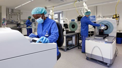
PET/MRI scanners unite two medical imaging techniques, but fully capitalising on the combination’s potential has proven difficult. Now, however, a team led by André Martins at the Werner Siemens Imaging Center (WSIC) in Tübingen have developed a new hybrid contrast agent that brings the best of both imaging worlds together—a significant step towards unlocking the full diagnostic capabilities of PET/MRI technology.
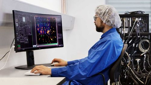
Medical imaging techniques like magnetic resonance imaging (MRI) and positron emission tomography (PET) are standard—and indispensable—tools in modern medicine. While MRIs furnish detailed visualisations of the structure and physiology of muscles, ligaments, tendons and inner organs, PET scanners enable tracking of single cells inside the body, on particular pathologically altered cells. A combination of the two imaging techniques offers enormous advantages, as just one examination is needed to pinpoint health issues with unprecedented precision.
The first PET/MRI hybrid systems were developed some twenty years ago, and Bernd Pichler, director of the Werner Siemens Imaging Center in Tübingen, pioneered their development. However, the technology’s full potential is still unfolding, which is partly because of the different contrast agents used to render the structures and functions in a body visible: MRI contrast agents are generally based on the heavy metal gadolinium, whereas radioactive fluorine-18 is frequently used for a PET signal.

Dual properties, one molecule
“Combining two different contrast agents is no simple task,” says André Martins, group leader at WSIC and professor of advanced preclinical metabolic imaging and cell engineering at the Eberhard Karl University of Tübingen. The main problem is that MRI scans require up to a billion more contrast molecules than a PET scan. Now, a research team from Tübingen and Prague led by Martins have found a solution to the problem: after years of intense effort, they have developed a multipurpose contrast molecule containing both gadolinium and radioactive fluorine-18.
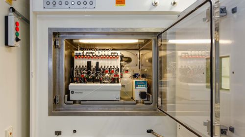
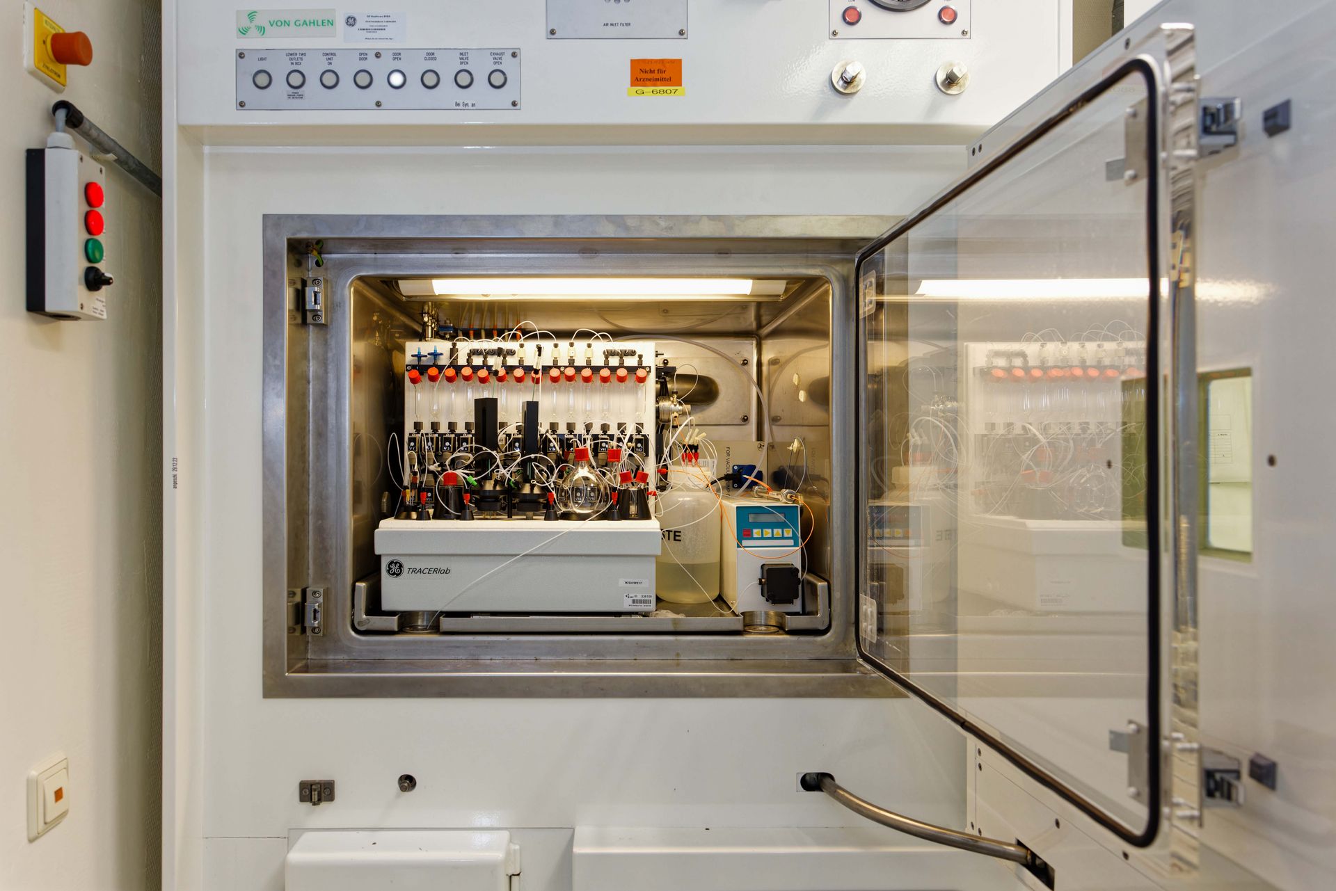
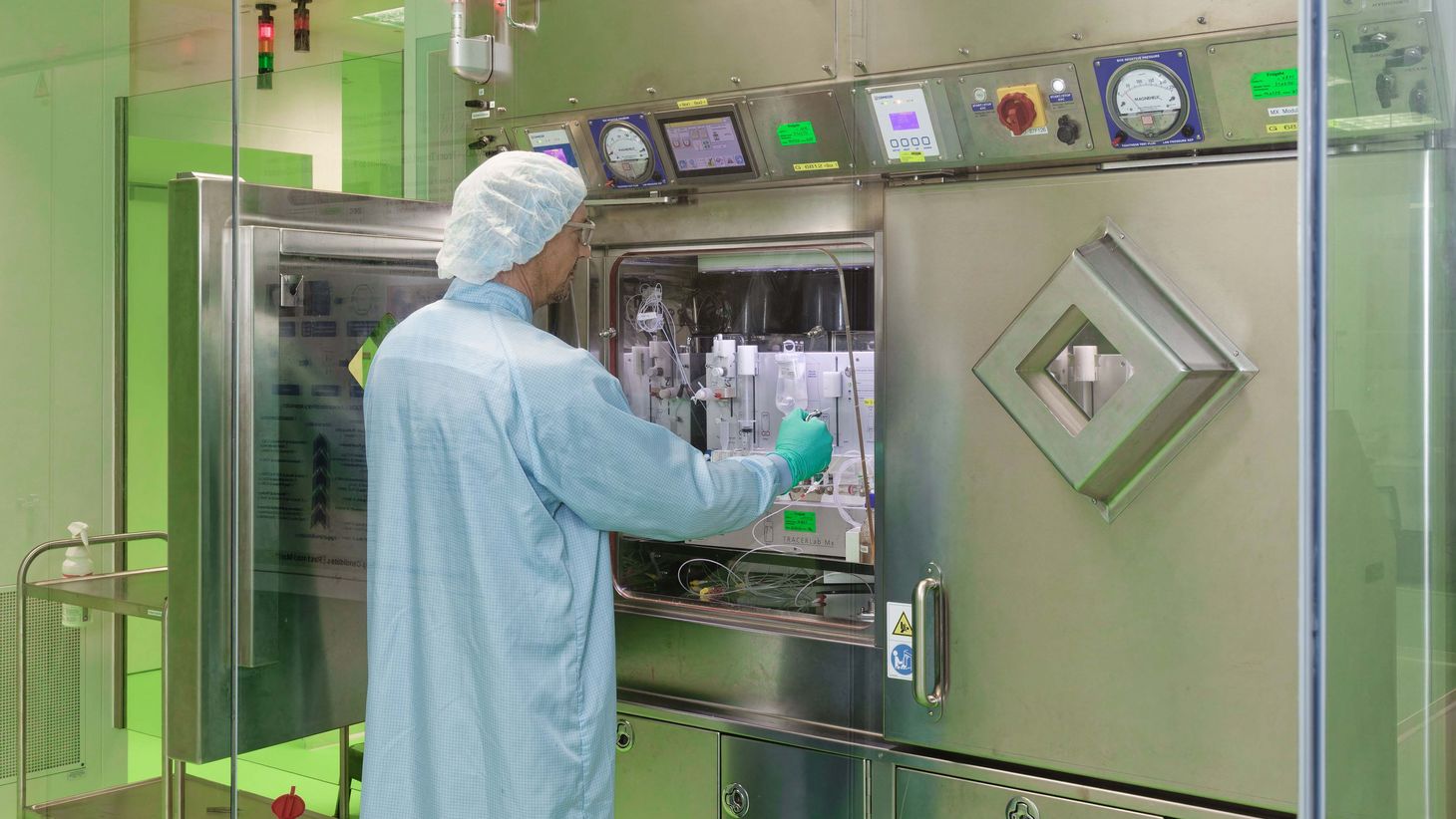
The researchers recently published an article about this groundbreaking innovation in the journal Angewandte Chemie International Edition.(*) In the article, they describe how to develop a gadolinium-based molecule that contains non-radioactive fluorine-19 atoms. At the final stage of synthesis, some of these fluorine-19 atoms are chemically replaced with radioactive fluorine-18, allowing the molecule to serve as a dual-purpose agent, effective in both MRI and PET scans.
The procedure is fast and precise: it took less than thirty minutes for the researchers to produce enough of the agent to examine five test subjects in preclinical models. Another advantage of the molecule is that it remains stable inside the body, which is an essential requirement for clinical use. In addition, it carries all the properties of current MRI contrast agents but is enriched with the added functionality of a PET signal, enabling simultaneous MRI and PET imaging for more detailed results. “The molecule opens up completely new possibilities in precision medicine,” André Martins says.
Good for patients
Patients benefit from combined PET/MRI examinations. In general, imaging procedures take quite a bit of time, but the combined examination means one less appointment with the doctor and one hour less spent inside the scanner. However, as Martins says, the real breakthrough lies in the enhanced precision and entirely new diagnostic insights enabled by the new molecule at the heart of this technology.
In their study, the researchers already presented the first positive diagnostic outcomes. “We first tested the contrast agent’s safety in healthy mice,” Martins explained. “Thanks to the new molecule, we found early indicators of kidney failure in two of seven mice.” The researchers made this observation because, counterintuitively, damaged kidneys displayed more heterogeneous gadolinium contrast agent elimination—sometimes even clearing it faster—than healthy organs.
While conventional MRI can indicate kidney abnormalities, it may miss subtle accumulations and inflammation sites. This is where the PET signal in the newly developed molecule comes in: it enables the researchers to identify the precise amount of the accumulated contrast agent and to locate accurately small inflammations—such as the affected kidneys in the observed mice. These details can easily be overlooked when relying solely on MRI.
More precise localisation of metastases
In the long run, the researchers believe the newly developed molecule will significantly enhance the ability to recognise and characterise cancer cells. Martins, the lead researcher, says that his team have now begun studies with tumour models. “Although the data are still preliminary, we may be able to localise metabolic processes in a tumour’s microenvironment much more precisely than with previous methods.” Additionally, Martins explains that this new approach could help determine whether a tumour exhibits necrosis, contains dead cells or is metabolically inactive. The advancements in imaging techniques will also aid in planning surgical interventions, ensuring that doctors can accurately target the right tissue when performing biopsies.
André Martins also believes it will be possible to use the new method to create additional contrast agents that are designed to target relevant alterations in physiology—measuring the body’s pH values, for example. This is particularly important because some tumours have a lower pH than the surrounding tissue, yet, as Martins explains, there’s currently no existing method to monitor pH levels within the body.
The researchers have already patented their new PET/MRI hybrid contrast agent molecule and are now seeking potential investors to ensure that their innovation attains clinical application as quickly as possible.
> (*) Link to the study

