Sophisticated implants
Fabricating made-to-measure implants with a 3D printer is one of the central components of the MIRACLE II project at the University of Basel. And now, researchers in the team are taking the first steps towards printing implants that can be reabsorbed by the body—potentially doing away with the need for follow-up surgeries.
Gentle, minimally invasive and highly precise bone surgery: this is the aim of researchers in the MIRACLE II project at the University of Basel and the University Hospital Basel. In the project—which receives funding from the Werner Siemens Foundation—the interdisciplinary team are developing a robot-guided surgical system that uses a laser to cut bones. And at the hospital’s inhouse 3D-printing lab, they’re fabricating implants that fit precisely into the pre-cut bones.
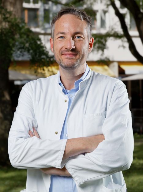
Last year, the team led by Professor Florian Thieringer, senior physician for oral and cranio-maxillofacial surgery and head of the inhouse 3D printing lab at the University Hospital Basel, successfully implanted an artificial calvaria (skull cap) designed and printed at the hospital in a patient—a first in Europe. In order to comply with the requirements of Switzerland’s Medical Devices Ordinance, they founded the start-up PoC-APP with the purpose of providing regulatory support to the hospital and passing on the know-how gained to other hospitals. Currently, the implants printed in Basel are made of plastics or bone substitutes; in future, however, the researchers are hoping to fabricate implants using materials whose components can be completely reabsorbed in vivo and replaced by body tissue that grows naturally.
In a feasibility study* recently published in 3D Printing in Medicine, a team led by Thieringer tested various patient-specific implants made of this kind of bioresorbable material—specifically, a mixture of lactic acid and tricalcium phosphate that has already been approved for use in humans.
Plates, meshes, bone scaffolding
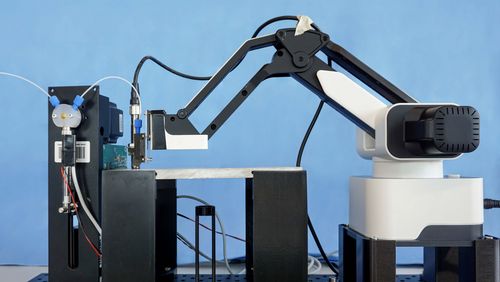
For the study, the researchers adopted the same basic procedure as for their standard 3D-printed implants. First, they used computer tomography to generate pictures of the damaged body part; the images then served to create a digital template for a patient-specific implant that was printed using a special Arburg 3D printer. The freshly fabricated implant was placed in an ultrasonic bath to remove even the tiniest of impurities and, in a final step, sterilised.
The researchers demonstrated that this method can be used to produce an entire range of different bioresorbable implants. Examples include the following: plates to fixate broken bones; various types of implants for orbital floor (eye socket) fractures; so-called meshes that facilitate jawbone regeneration; and bone scaffolds that are permeated with pores.
The bioresorbable 3D implants have not yet been approved for use in patients. “But,” Florian Thieringer says, that’s our next step.” He adds, “We’re currently working on validating the processes so that we can use them in clinical practice.” The researchers are familiar with the regulatory procedures, as they’ve already gone through the steps to gain approval for the 3D-printing and clinical application of their implant made of polyether ether ketone (PEEK), the material used in the plate for last year’s calvaria implant.
One surgery less
The fact that bioresorbable materials disintegrate inside the body over time is of great advantage in certain situations. As Florian Thieringer explains, “The idea is that the body’s own tissue will at some point replace the polymers.” One clear benefit is that follow-up operations to remove a non-resorbable implant may no longer be necessary.
Florian Thieringer says a specific application for the new technology would be for bone augmentation procedures that are frequently conducted before a patient receives a dental implant. If several teeth have already fallen out, deterioration in the underlying jawbone is not uncommon; in such cases, the dental surgeon will first work to rebuild the bone before inserting the implant. “When we use materials like titanium meshes, we have to remove them at a later stage. So, we always need to schedule another surgery—and that’s hard on patients,” Thieringer explains.
The bioresorbable mesh could also benefit children who require surgery to repair birth defects like cleft palates or skull deformities. “In current practice, the bones in the cleft area are augmented or the cranial bones are remodelled. The problem is that children are still growing—rigid materials are unsuitable because they influence growth patterns,” Thieringer says. By contrast, materials that are absorbed into the body would exert no such exterior pressure.
Added value at no added cost?
The researchers still need to master several challenges before the resorbable materials from the 3D printer are ready for use in clinical practice. For example, it isn’t clear which materials are best for which purposes. Thieringer says that magnesium is an interesting candidate: “It’s now been tested in initial applications and ongoing projects with a cautious degree of success.” However, clinical trials in humans are also necessary to ensure general safety, including that of the material’s resorption behaviour. When magnesium dissolves, it creates hydrogen, and, as Florian Thieringer explains, “this causes gas to form around the implants, which can have negative effects on bone healing”.
Choosing the best material as well as determining its processing and its biological tolerability in the human body represent major hurdles that the researchers have to take on their way to incorporating resorbable materials in surgical interventions. Other problem points are technologies and production processes, while still another issue concerns regulatory specifications. Regarding the latter, Thieringer points out that “the entire treatment chain has to be validated and approved—from medical imaging and planning, on to printing and sterilising the implants, all the way to introducing them into a patient’s body”. And last but not least are aspects regarding economic viability: “We want to create added value for our patients without generating significant added costs.”
Thieringer is convinced that the new, resorbable materials are the way of the future. And he’s not alone. Recently, his 3D printing lab welcomed a new family member, as it were—a 3D printer the size of an armoire. “The Arburg company was so impressed by our research that they’re allowing us to use their flagship 3D printer,” Thieringer says. The elite model can print several components at the same time—a water-soluble scaffold and a bone replacement, for example. All in all, highly auspicious signs for 3D printing in medical interventions.
> * Link to the study